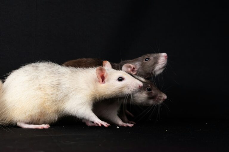
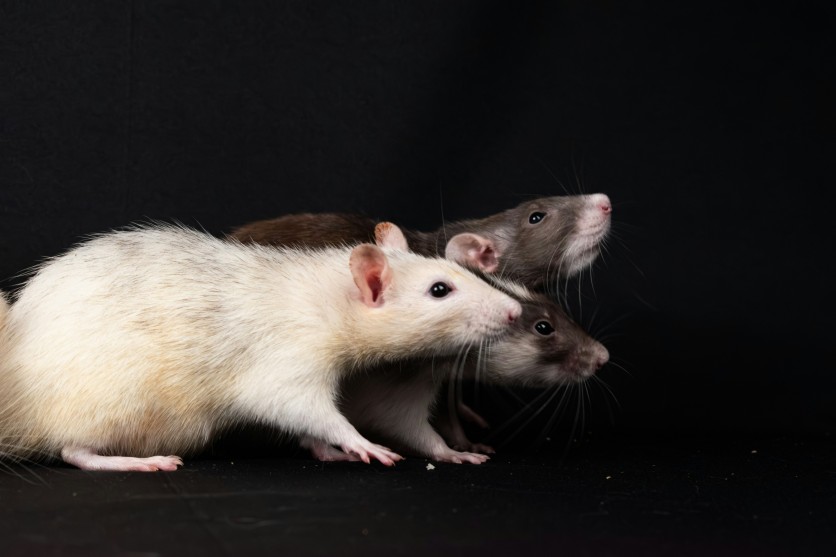
For decades, sports scientists have focused on how excessive training damages muscle tissue, strains tendons, and suppresses the immune system. A study published today in the journal Neuroscience shifts attention to a less-examined organ: the brain. Researchers at the University of São Paulo (USP) and the University of Campinas (Unicamp) in Brazil report that chronic overtraining disrupts dopamine homeostasis in hippocampus of mice — a brain region central to memory and emotional regulation — and that this disruption persists well into the recovery period, 48 hours after exercise has ended. The system, the researchers found, simply cannot shut off.
The finding matters for a straightforward reason: millions of competitive and amateur endurance athletes worldwide experience overtraining syndrome (OTS) during their careers — estimated among competitive athletes over career at 20 to 60 percent — and a significant share of those athletes report not just physical collapse but cognitive and psychological ones: anxiety, sleep disruption, inability to concentrate. Until now, those symptoms had no clearly demonstrated neurochemical home.
Overtraining Syndrome Has a Documented Psychological Toll — and No Validated Biomarker
Overtraining syndrome arises when training volume and intensity persistently outpace an athlete’s capacity for recovery, triggering a cascade of symptoms that extend well beyond muscle fatigue. Elite endurance athletes — long-distance runners, cyclists, triathletes — are considered especially at risk, but the population of at-risk athletes has expanded as high-intensity fitness culture has moved into mainstream amateur sport.
What has long puzzled clinicians is why overtrained athletes so frequently report psychological and cognitive symptoms alongside the physical ones: anxiety, sleep disturbance, mood disruption, difficulty concentrating. Prior research established that the central nervous system is involved. A landmark 2019 study published in Current Biology found that overtrained endurance athletes showed reduced lateral prefrontal cortex activation and were more prone to impulsive, short-term-reward-seeking decisions — an effect that mirrored what researchers had previously observed from cognitive overwork. That study, conducted at the Hôpital de la Pitié-Salpêtrière in Paris, was the first to demonstrate that physical training overload could produce a neurological fatigue comparable to mental exhaustion.
What it did not establish was the specific neurochemical mechanism. The new Brazilian study targets that question directly.
What the Experiment Did
Lead author Alisson Luiz da Rocha — whose doctoral work was conducted at USP’s School of Physical Education and Sport of Ribeirão Preto (EEFERP-USP), with subsequent postdoctoral research at Unicamp and a research fellowship at the University of Michigan — divided laboratory mice into groups for the experiment.
The first group underwent a single acute exercise session: each animal ran on a treadmill until reaching exhaustion, simulating the kind of intense one-off effort a recreational athlete might experience before an event.
The second group followed an eight-week chronic overtraining protocol, designed to mirror the physiological profile seen in elite athletes with OTS. Training loads were progressively increased over eight weeks. In the final week, the intensity escalated: animals completed two treadmill sessions per day, separated by only four hours of recovery time — a deliberately insufficient rest window. A performance test concluded the protocol.
After exercise, the researchers analyzed hippocampal tissue from both groups, measuring dopamine and key proteins: tyrosine hydroxylase (TyrH), the enzyme that catalyzes the rate-limiting step in dopamine synthesis, and vesicular monoamine transporter 2 (VMAT2), which packages dopamine for synaptic release.
Dopamine Stayed High — and the Serotonin Hypothesis Did Not Hold Up
The acute exercise group showed what might be expected: dopamine levels in the hippocampus rose immediately after the treadmill run, consistent with exercise’s well-established stimulatory effects on the brain. This spike is a normal adaptive dopamine response to exercise.
The findings for the chronic overtraining group were different. Forty-eight hours after the final performance test — during what should have been a recovery phase — dopamine and TyrH remained elevated significantly in hippocampal tissue. The dopaminergic system had not returned to baseline. The brain’s machinery for synthesizing dopamine was still running at high capacity even as the animals were supposed to be recovering.
The serotonin finding is theoretically at least as significant as the dopamine one. Serotonin levels showed no statistically significant changes in either group. This directly challenges the OTS research toward serotonergic mechanisms that the serotonin fatigue hypothesis — first proposed by biochemist Eric Newsholme in the 1980s and influential for decades — had oriented. The new data suggest the hippocampal dopaminergic system may carry a specific and disproportionate burden under overtraining conditions, and that decades of OTS research oriented toward serotonergic mechanisms may need to be reconsidered.
Study coordinator Prof. Adelino Sanchez Ramos da Silva of EEFERP-USP noted that these neurochemical changes, if they occur similarly in humans, could explain sleep and anxiety symptoms commonly reported by overtrained athletes. Persistently elevated dopamine during rest — a time when the brain normally downregulates arousal-promoting signals — is inconsistent with healthy sleep architecture and emotional regulation.
The study did not directly measure sleep quality, alertness, or anxiety in the animal subjects. The connection between the observed dopamine elevation and these clinical symptoms in humans remains inferential, and the research team explicitly flagged this as a question for future investigation.
Why the Hippocampus, and How the Dopamine Pathway Works
Dopamine is frequently described as a “pleasure chemical,” but its functions are considerably more specific. In the hippocampus, dopamine plays important roles in memory consolidation, spatial navigation, and the encoding of novel experiences. Two dopamine-producing regions project into the hippocampus: the locus coeruleus (LC) and the ventral tegmental area (VTA). Prior research has shown that even light exercise stimulates hippocampal dopamine neurons, boosting learning and memory in the short term.
The problem the Brazilian study identifies is one of timing and proportion. Dopamine is not harmful; the authors are careful to emphasize this. What becomes harmful is the loss of homeostatic regulation — when dopamine remains chronically elevated during periods when the brain should be in a lower-arousal, restorative state.
Understanding why requires a brief look at the catecholamine biosynthesis pathway. Dopamine is synthesized through a two-step process: the amino acid L-tyrosine is first converted to L-DOPA by tyrosine hydroxylase (TyrH), and L-DOPA is then converted to dopamine by aromatic L-amino acid decarboxylase (AADC). TyrH is the rate-limiting enzyme — the step that controls how much dopamine can be made. Once synthesized, dopamine is packaged into synaptic vesicles by VMAT2 for release into the synapse.
The persistence of elevated TyrH 48 hours after exercise cessation is what makes this study mechanistically significant. TyrH being elevated is not an incidental finding — it means the signaling pathway that drives dopamine production has been durably altered, not merely temporarily activated. The machinery is still making dopamine at high rates when it should be idling. This makes TyrH a potential neurochemical biomarker of overtraining syndrome in preclinical models — a finding the study authors explicitly highlight.
Complementary Evidence: BDNF Decline Adds to the Picture
Independent corroboration comes from a 2025 study in Nutrients, conducted by a separate research group at Ariel University in Israel, which found complementary evidence in a resistance-training overtraining mouse model. That study found overtraining was associated with elevated D1R and declining BDNF — alongside significant declines in brain-derived neurotrophic factor (BDNF), a protein critical for hippocampal neuroplasticity and adult neurogenesis — and documented spatial memory deficits as consequence.
Together, the two studies describe an emerging pattern: chronic excessive exercise disrupts the hippocampal dopaminergic system in multiple, complementary ways. In the Brazilian model, the synthesis machinery runs unabated during recovery. In the Israeli model, receptor expression is dysregulated and neurotrophic support declines. The common thread is a hippocampus whose plasticity and homeostatic regulation have been compromised by training loads that exceeded its capacity to adapt.
This matters for athletes because BDNF is the protein most reliably increased by moderate aerobic exercise — it is one of the primary reasons exercise is recommended for cognitive health and depression prevention. The inverted-U relationship between exercise dose and brain health is becoming clearer: moderate exercise benefits the hippocampus; chronic overtraining may damage it.
What Does This Mean for Athletes and for Diagnosis?
As first author da Rocha told FAPESP’s science news service, the findings reinforce the view that physical activity impacts the brain far beyond muscle adaptations. For competitive athletes and coaches, the neurochemical framing redefines what recovery is and why it is required — not merely as tissue repair but as an active neurochemical reset.
For clinicians, the TyrH finding offers something more specific: a potential biomarker direction. Current detection of overtraining syndrome is largely behavioral and performance-based — athletes see their race times worsen, their mood deteriorate, their motivation collapse — with no validated neurochemical marker exists for reliable early detection. A reliable neuroendocrine or neurotransmitter-based signal that could be detected before full OTS develops would represent a significant clinical advance. The research remains in preclinical animal models, and the path from a mouse hippocampus finding to a clinically deployable human biomarker is long and uncertain. But the direction is now testable.
For the broader public of amateur athletes — the millions of gym-goers and weekend competitors who train intensively without the support infrastructure of elite sport — the practical implication is straightforward: the biological cost of insufficient recovery is not purely muscular. The brain, too, is subject to a dose-response curve. More is not always better.
What Comes Next
The team at USP and Unicamp is already working on a new international collaboration aimed at identifying the earliest biological signs of deviation from the beneficial adaptations that regular exercise produces FAPESP science news service reports. The goal is to identify, earlier and more precisely, the transition from healthy training stress to pathological overtraining — a threshold that remains notoriously difficult to locate. Co-investigators Ana Paula Pinto and João Pedro Floriano, both based at EEFERP-USP, are also involved in ongoing work building on the study’s findings.
The serotonin null finding may prove to be a turning point in how that search is oriented. If the dopaminergic system — not the serotonergic one — is the primary hippocampal casualty of overtraining, then future diagnostic targets, intervention strategies, and recovery protocols designed around serotonin may be looking in the wrong place.
Frequently Asked Questions
What specifically happens to the brain during overtraining syndrome?
The new study finds that chronic overtraining disrupts dopamine homeostasis in the hippocampus — the brain region governing memory, spatial navigation, and emotional regulation. Specifically, both dopamine and tyrosine hydroxylase (the rate-limiting enzyme in dopamine production) remain elevated 48 hours into the recovery period, long after exercise has ended. The brain’s dopamine synthesis machinery does not return to baseline when it should. This persistent elevation, during a period when the brain is supposed to be in a lower-arousal restorative state, is inconsistent with healthy sleep architecture and emotional regulation, and may explain why overtrained athletes frequently report anxiety, sleep disruption, and cognitive difficulties alongside physical symptoms.
Does overtraining also affect serotonin?
The study found no statistically significant changes in serotonin levels in either the acutely exercised or the chronically overtrained mice — a finding that directly challenges the longstanding serotonin fatigue hypothesis, which has attributed chronic exercise fatigue primarily to rising brain serotonin since it was first proposed in the 1980s. The data suggest that hippocampal dopamine, not serotonin, may be the primary neurochemical casualty of overtraining, which has potential implications for how OTS research and clinical interventions are oriented going forward.
Can overtraining syndrome be measured with a blood test or biomarker?
Not yet — there is currently no widely validated biochemical or neurochemical marker for OTS, and the new finding remains in a preclinical mouse model. The study identifies tyrosine hydroxylase as a potential biomarker candidate in animal models, given that its persistent elevation distinguishes the chronically overtrained brain from the acutely exercised one. The path from a mouse hippocampus finding to a clinically deployable human biomarker is long and requires human validation studies. The research does, however, establish a testable direction that did not clearly exist before.
How much exercise is too much — and how would I know if I’m overtrained?
No universal threshold exists; individual response varies substantially by fitness level, recovery quality, nutrition, sleep, and psychological stress. OTS develops on a continuum: functional overreaching (recovers in days to weeks), non-functional overreaching (recovers over weeks to months), and full overtraining syndrome (may require months to years). Warning signs that go beyond normal fatigue include persistent performance decline despite rest, psychological symptoms (anxiety, mood disruption, loss of motivation), sleep disturbance, and unusually frequent illness. The neurochemical research suggests that the brain’s failure to reset during rest periods — not merely the training volume itself — is a key feature of OTS, which is why adequate recovery time, not just reduced training load, is the primary intervention.
The paper “Excessive training alters hippocampal tyrosine hydroxylase and dopamine homeostasis in mice” is published in Neuroscience is available at ibroneuroscience.org (paywalled). The research was funded by FAPESP (São Paulo Research Foundation).